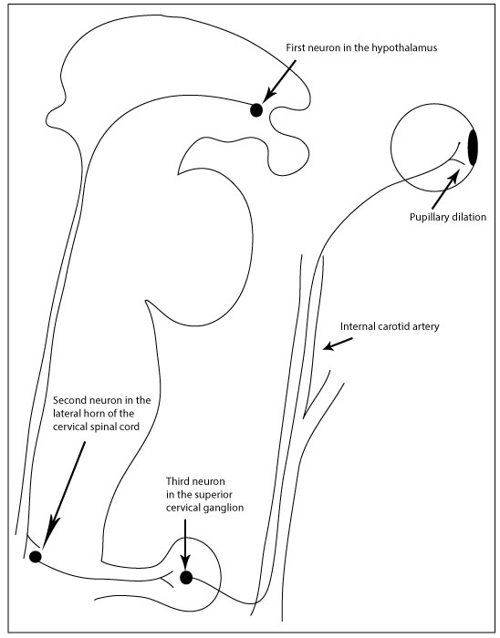
The pupillary dark reflex consists of bilateral pupil dilation, which is also called mydriasis, to dark. Some neurons in the hypothalamus, which is in the inferior cerebrum, receive information about light and dark from the eyes, probably from both sides, so that there is an equal bilateral efferent response to unilateral afferent stimuli, similar to the pupillary light reflex. The three neuron chain that makes up the efferent limb of this reflex is all ipsilateral. The first neuron projects from the hypothalamus down the brainstem and the lateral column of the upper spinal cord to synapse on a preganglionic sympathetic neuron in the lateral gray horn. This second neuron projects through nerves in the chest to synapse on the postganglionic sympathetic neuron in a ganglion in the neck. This third neuron projects through small sympathetic nerves that ascend along the walls of arteries. They then travel in cranial nerve branches into the orbit to synapse on the iris dilator muscle, which is smooth muscle, to cause pupil dilation.
Unilateral dysfunction of the efferent limb of the pupillary dark reflex causes anisocoria. The anisocoria becomes better in the light, because pupillary constriction is normal bilaterally. The anisocoria becomes worse in the dark because pupillary dilation is normal on the normal side.
Next:
